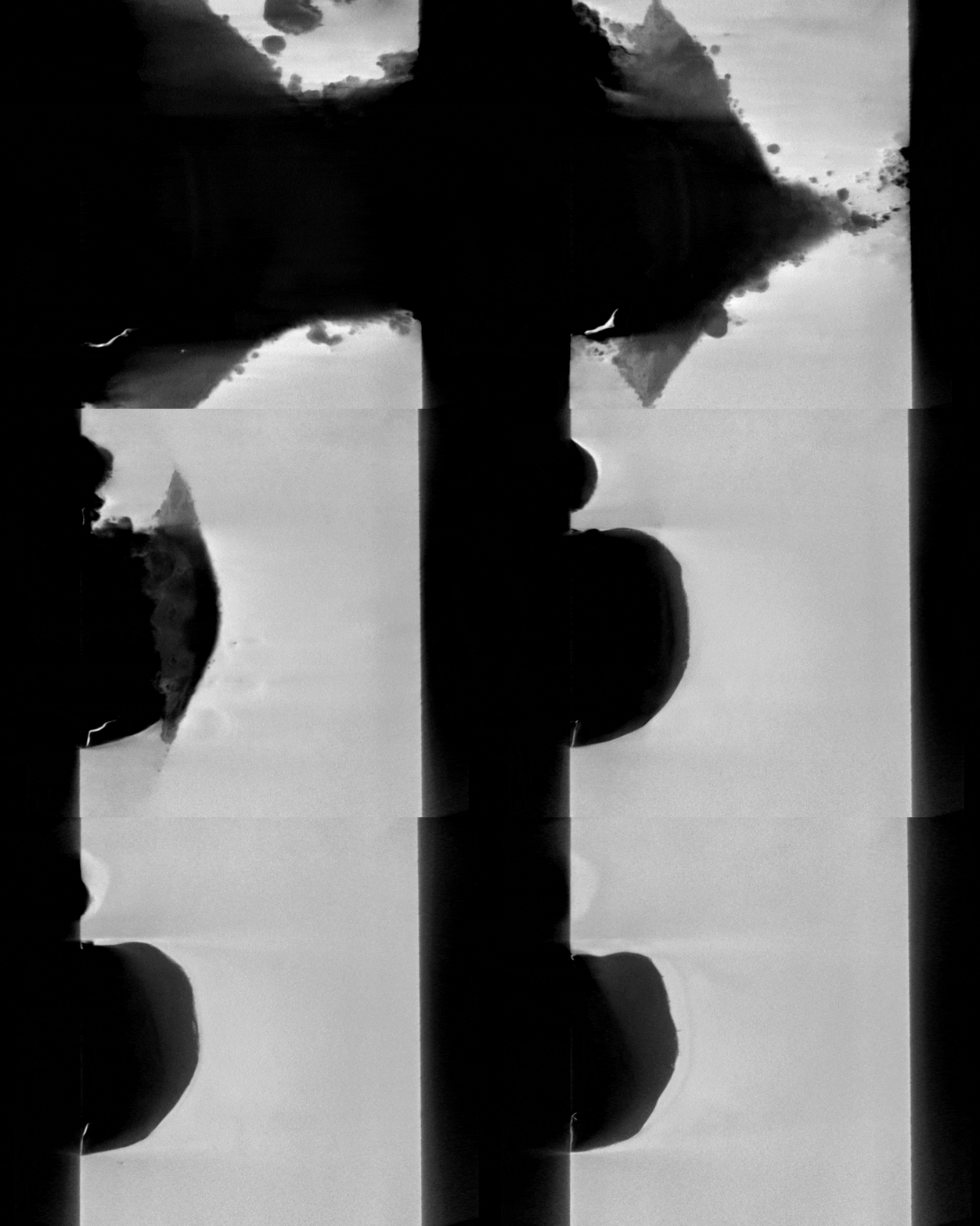
Xradia Instructions: Difference between revisions
Jump to navigation
Jump to search
Created page with "=User Tutorials= *'''Xradia Versa User’s Guide''' *Media:Zeiss-xrm-xradia-520-versa-advanced-training-selected-topics_reve.pdf|'''Xradi..." |
|||
| (7 intermediate revisions by the same user not shown) | |||
| Line 7: | Line 7: | ||
=Scanning= | =Scanning= | ||
*[[Media:updatedzeissxradia5xxversa_scout-and-scan_quickref.pdf|'''Scout-and-Scan™ Control System Quick Reference Guide''']] | *Quick Reference Guide: [[Media:updatedzeissxradia5xxversa_scout-and-scan_quickref.pdf|'''Scout-and-Scan™ Control System Quick Reference Guide''']] | ||
*[[Media:Zeissxrm_technical-procedure-scout_and_zoom-version-10 | *Log Sheets: [[Media:Xradia_log_sheet_general.pdf|'''PDF Log sheets''']] and [[Media:Xradia_log_sheet_general.xlsx|'''Excel Log sheets''']] | ||
*[[Media: | |||
==Interior Tomography== | |||
*[[Media:Zeissxrm_technical-procedure-scout_and_zoom-version-10.pdf|'''Scout-and-Zoom for High Resolution Interior Tomography v10''']] | |||
==Vertical Stitching== | |||
*[[Media:ZEISSXRM_QuickReferenceGuide_VerticalStitching_v11.pdf|'''Quick Reference Guide for Vertical Stitching v11''']] | |||
*[[Media:ZEISSXRM_TechnicalProcedure_VerticalStitching_v11.pdf|'''Technical Procedure for Vertical Stitching v11''']] | |||
==High Aspect Ratio Tomography (HART)== | |||
*[[Media:034zeiss-xrm-technical-procedure-high-aspect-ratio-tomography-hart.pdf|'''High Aspect Ratio Tomography (HART) v10.6''']] | |||
| Line 15: | Line 27: | ||
=Reconstruction= | =Reconstruction= | ||
*[[Media:ZEISSXRM_Scout-and-Scan Reconstructor_QuickRefGuide.pdf|'''Reconstructor Quick Reference Guide v11''']] | |||
*[[Media:ZEISSXRM_Scout-and-Scan Reconstructor_Manual.pdf|'''Reconstructor Manual v11''']] | |||
| Line 20: | Line 34: | ||
=Special Topics= | =Special Topics= | ||
==Calibration== | |||
*[[Media:ZEISS_XRM_Technical_Procedure_-_CT_Scaling_Instructions_G000135.pdf|'''CT Scaling Calibration''']] | |||
==Image Artifacts== | |||
*[[Media:ZEISS_XRM_Technical_Procedure_-_Filtered_Secondary_Referencing_For_Ring_Artifact_Reduction_-_Xradia_520_Versa.pdf|'''Filtered Secondary Referencing for Ring Artifact Reduction''']] | |||
*[[Media:ZEISS XRM Technical Procedure - Despeckling Filter.pdf|'''Despeckling Filtering''']] | |||
[[Image:Redirect.jpg|15px]] [[#top|''Return to Contents'']] or [[Main_Page|''Return to Main Page'']] | [[Image:Redirect.jpg|15px]] [[#top|''Return to Contents'']] or [[Main_Page|''Return to Main Page'']] | ||
=Dual-Energy Imaging= | ==Dual-Energy Imaging== | ||
*[[Media:ZEISS_XRM_Xradia_520_Versa_--_Guide_to_DSCoVer.pdf|'''Guide to DSCoVer v10''']] | |||
*[[Media:Dualenergy_technical-procedure-zeiss_v4_draft_dis.pdf|'''Dual-Energy Imaging Manual''']] | |||
[[Image:Redirect.jpg|15px]] [[#top|''Return to Contents'']] or [[Main_Page|''Return to Main Page'']] | [[Image:Redirect.jpg|15px]] [[#top|''Return to Contents'']] or [[Main_Page|''Return to Main Page'']] | ||
=Phase Contrast Imaging= | ==Phase Contrast Imaging== | ||
*[[Media:ZEISS_XRM_Technical_Procedure_-_Phase_Contrast.pdf|'''Phase Contrast Imaging Manual''']] | |||
[[Image:Redirect.jpg|15px]] [[#top|''Return to Contents'']] or [[Main_Page|''Return to Main Page'']] | |||
=Image Tours= | |||
==Industrial Materials== | |||
*Refer to [https://youtu.be/tT3dz4VrwfE '''MicroCT Image Tour YouTube Video'''] | |||
*Hover over a thumbnail and click on its overlaid description to see the animated GIF or a larger version of the image. | |||
<gallery mode="packed-hover" heights=220px caption="MicroCT Scans for Industrial Materials"> | |||
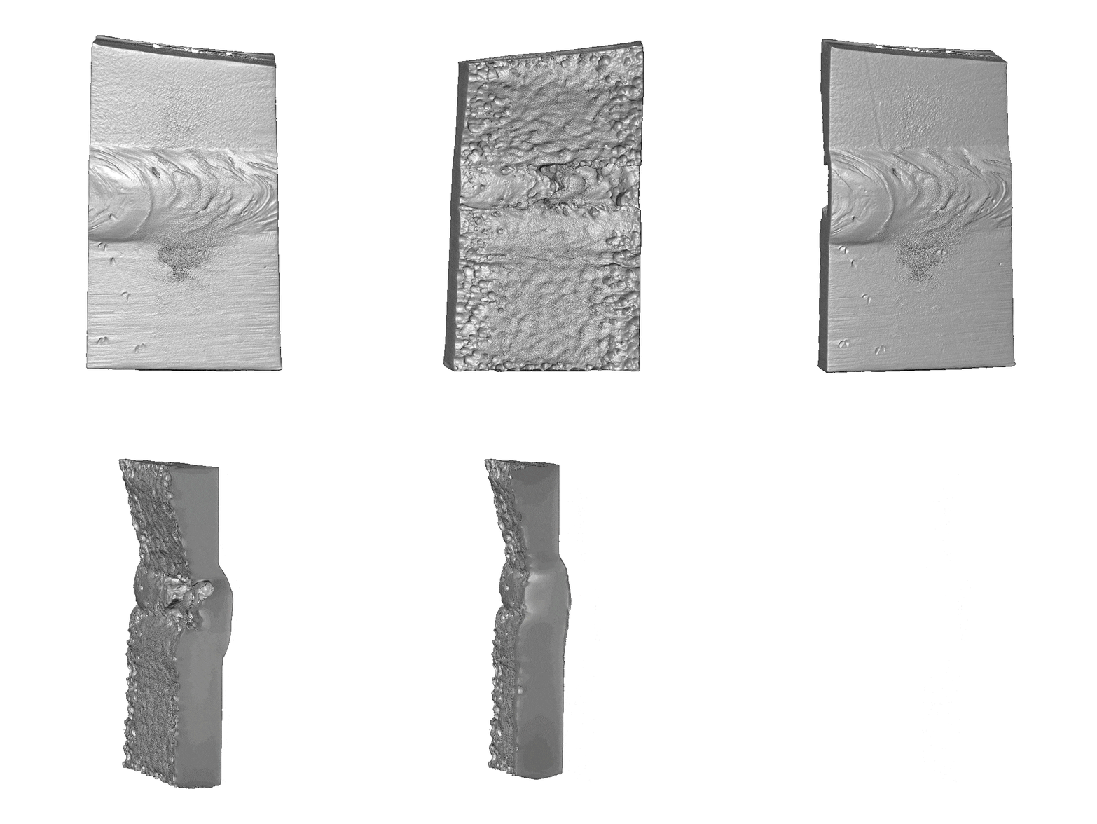
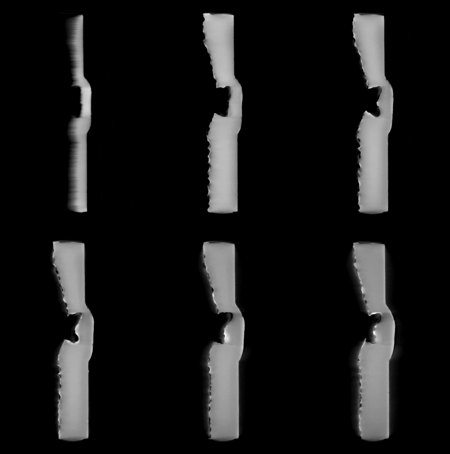
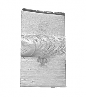
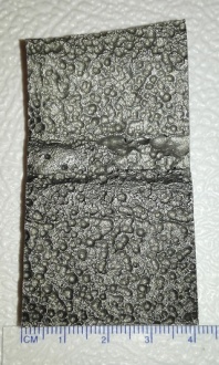
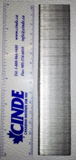
Image:002.0000_0.png|[[Media:002001-whitebg-movie_0.gif|Steel Welds Fusion Zone (50µm, 39 x 66 x 11 mm3)]] | |||
Image:Img_20170120_115108-copy_1.jpg|[[Media:002001-50um-xz_140-290.gif|Steel Welds Fusion Zone (50µm, 39 x 66 x 11 mm3)]] | |||
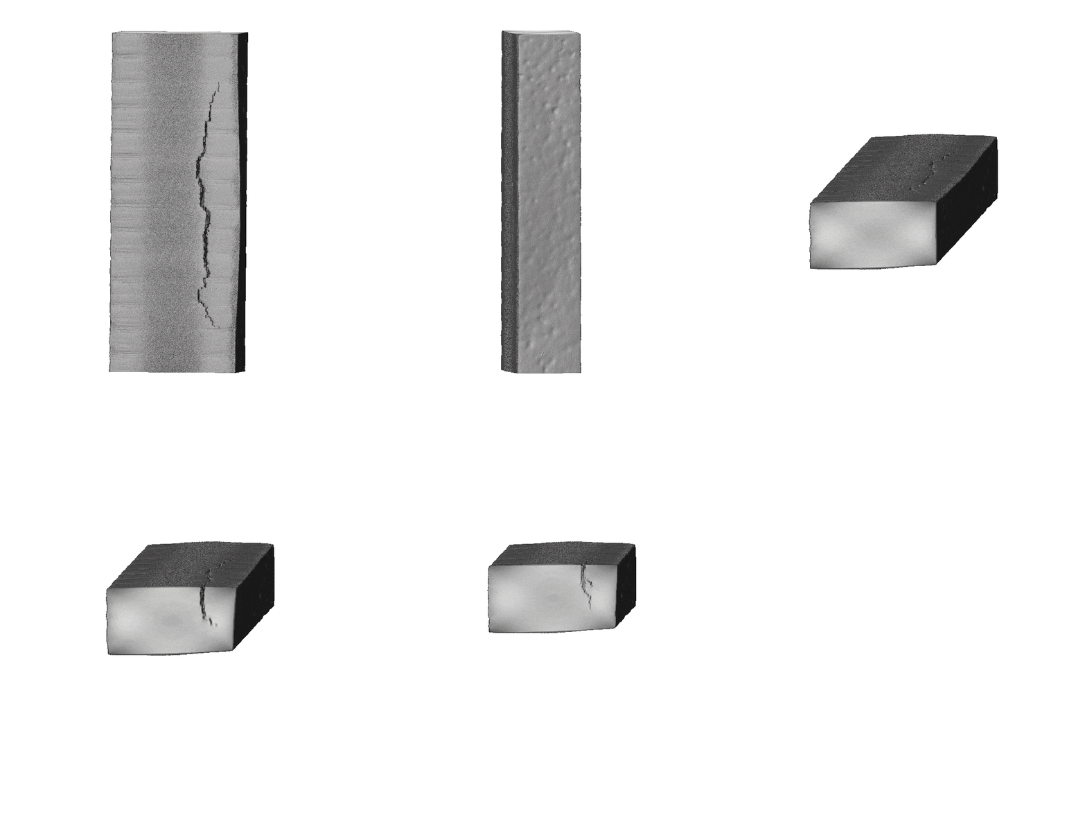
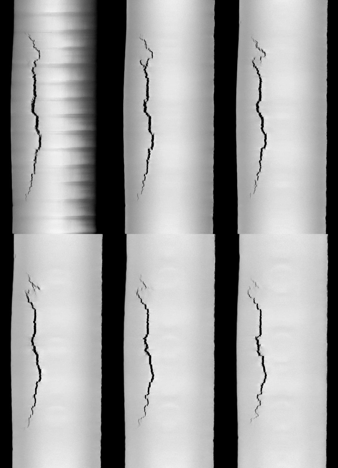
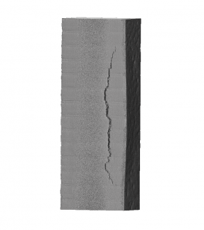
Image:003.0095.png|[[Media:003001-whitebg-movie.gif|Metal Crack (36µm, 137 x 28 x 15 mm3)]] | |||
Image:003001_microctwebsite_0.jpg|[[Media:003001-36um_cut_xz_310-390.gif|Metal Crack (36µm, 137 x 28 x 15 mm3)]] | |||
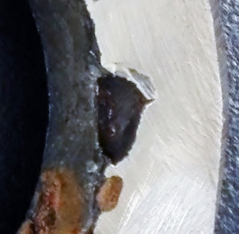
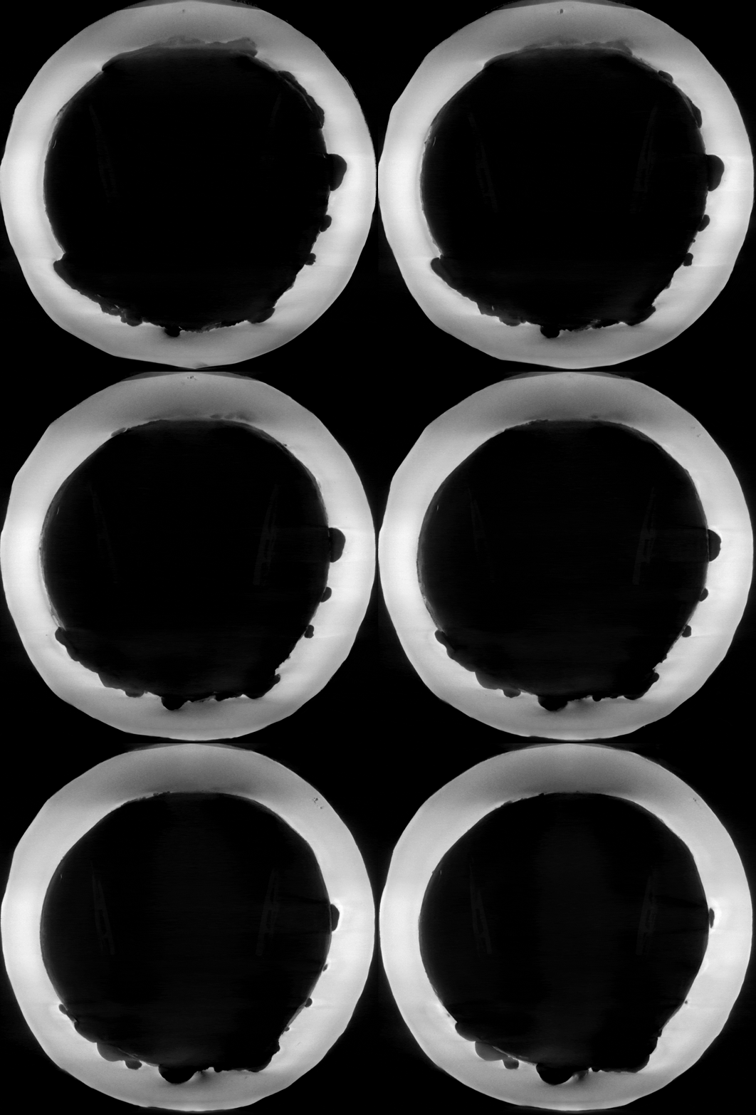
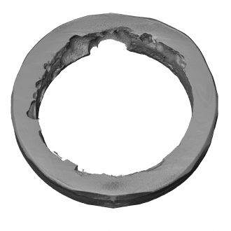
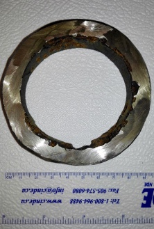
Image:001001.0000.png|[[Media:001001-whitebg-movie.gif|Corroded Welds Ring (53µm for full FOV, OD 93 mm x 14 mm thick)]] | |||
Image:001001_microctwebset.jpg|[[Media:001001_full_53um_cut_xz-60-120.gif|Corroded Welds Ring (53µm for full FOV, OD 93 mm x 14 mm thick)]] | |||
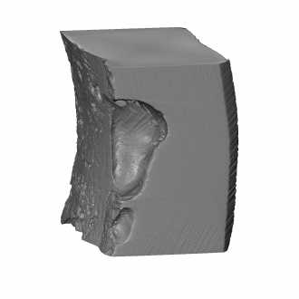
Image:Suncor001002.0000.png|[[Media:001002-whitebg-movie.gif|Corroded Welds Ring (21µm zoom in to right side)]] | |||
Image:001003_0.jpg|[[Media:001003_zoominside-21um_cut_xz-260-410.gif|Corroded Welds Ring (21µm zoom in to right side)]] | |||
</gallery> | |||
[[Image:Redirect.jpg|15px]] [[#top|''Return to Contents'']] or [[Main_Page|''Return to Main Page'']] | [[Image:Redirect.jpg|15px]] [[#top|''Return to Contents'']] or [[Main_Page|''Return to Main Page'']] | ||
Latest revision as of 20:02, 28 January 2021
User Tutorials
[edit]
 Return to Contents or Return to Main Page
Scanning
[edit]- Quick Reference Guide: Scout-and-Scan™ Control System Quick Reference Guide
- Log Sheets: PDF Log sheets and Excel Log sheets
Interior Tomography
[edit]
Vertical Stitching
[edit]
High Aspect Ratio Tomography (HART)
[edit]
Return to Contents or Return to Main Page
Reconstruction
[edit]
Return to Contents or Return to Main Page
Special Topics
[edit]Calibration
[edit]
Image Artifacts
[edit]
Return to Contents or Return to Main Page
Dual-Energy Imaging
[edit]
Return to Contents or Return to Main Page
Phase Contrast Imaging
[edit]
Return to Contents or Return to Main Page
Image Tours
[edit]Industrial Materials
[edit]- Refer to MicroCT Image Tour YouTube Video
- Hover over a thumbnail and click on its overlaid description to see the animated GIF or a larger version of the image.
- MicroCT Scans for Industrial Materials
-
-
-
-
-
-
-
-