Xradia Instructions: Difference between revisions
Jump to navigation
Jump to search
| Line 63: | Line 63: | ||
<gallery mode="packed-hover" heights=220px caption=" | <gallery mode="packed-hover" heights=220px caption="MicroCT Scans for Industrial Materials"> | ||
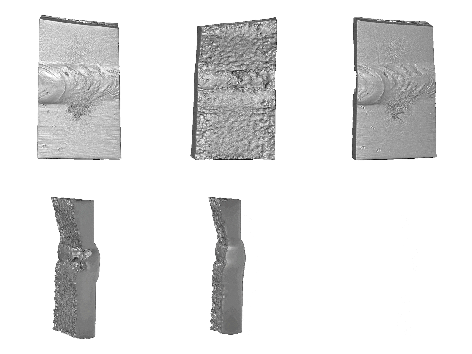
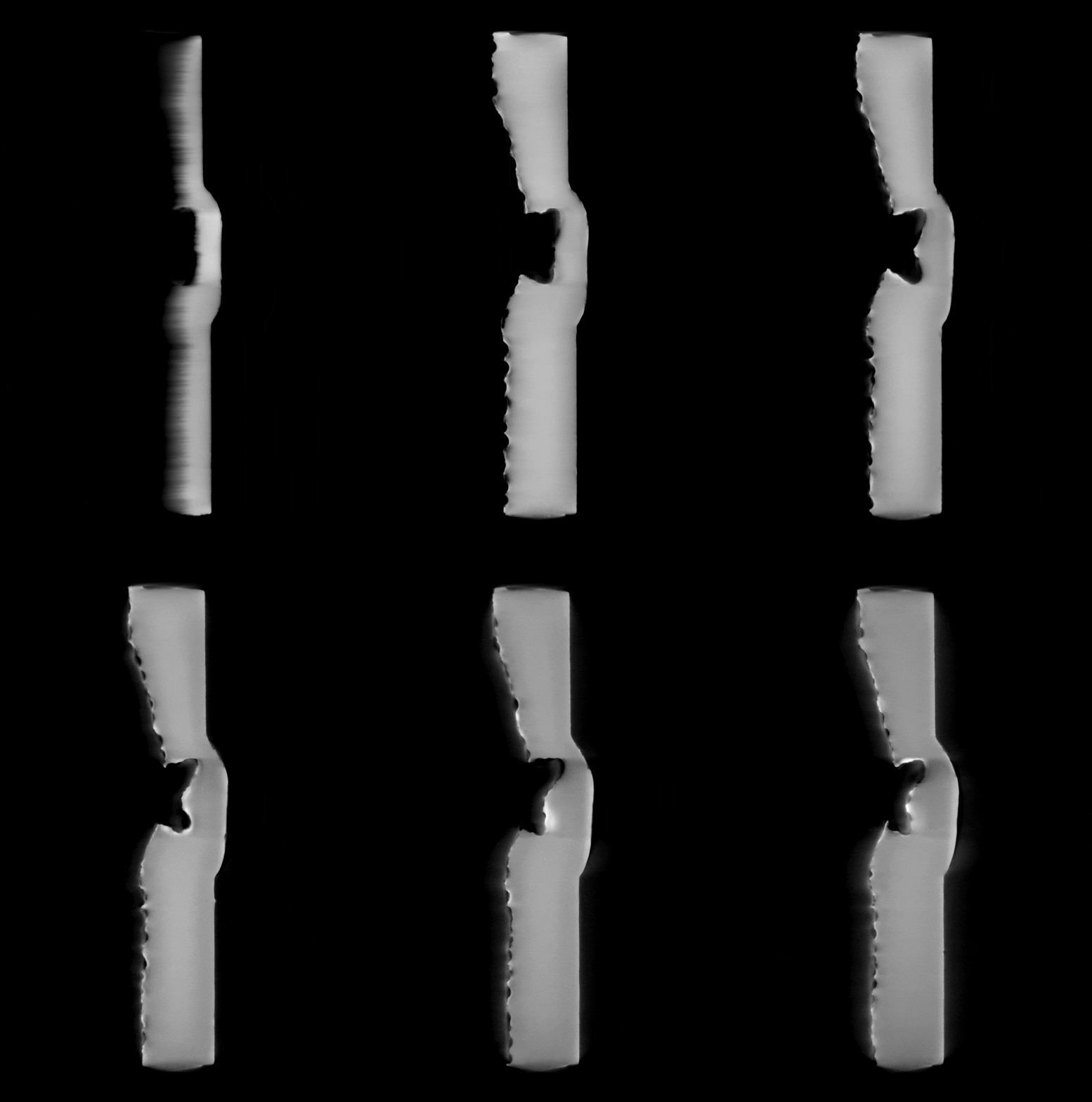
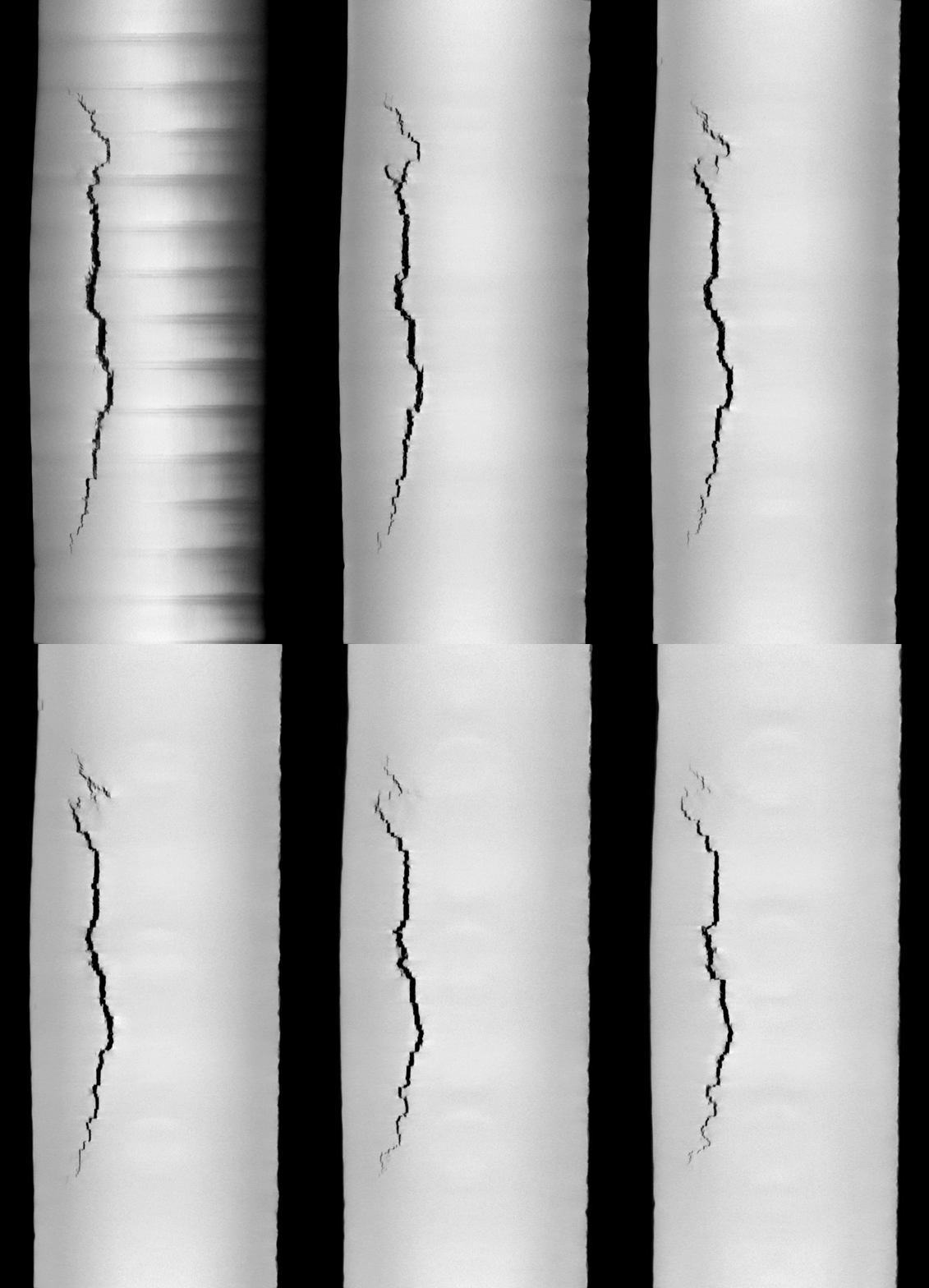
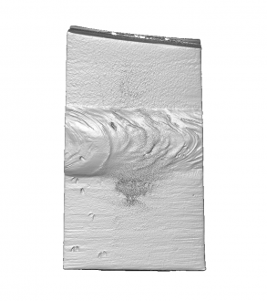
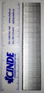
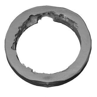
Image:002.0000_0.png|[[Media:002001-whitebg-movie_0.gif|Steel Welds Fusion Zone (50µm, 39 x 66 x 11 mm3)]] | Image:002.0000_0.png|[[Media:002001-whitebg-movie_0.gif|Steel Welds Fusion Zone (50µm, 39 x 66 x 11 mm3)]] | ||
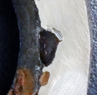
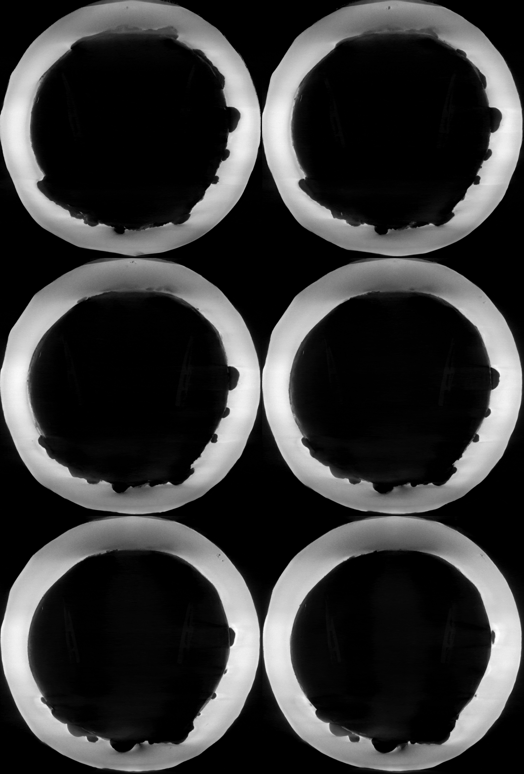
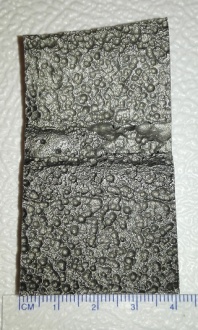
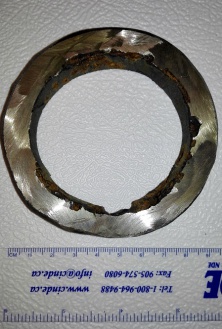
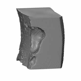
Image:Img_20170120_115108-copy_1.jpg|[[Media:002001-50um-xz_140-290.gif|Steel Welds Fusion Zone (50µm, 39 x 66 x 11 mm3)]] | Image:Img_20170120_115108-copy_1.jpg|[[Media:002001-50um-xz_140-290.gif|Steel Welds Fusion Zone (50µm, 39 x 66 x 11 mm3)]] | ||
Revision as of 23:57, 27 January 2021
User Tutorials
 Return to Contents or Return to Main Page
Scanning
- Quick Reference Guide: Scout-and-Scan™ Control System Quick Reference Guide
- Log Sheets: PDF Log sheets and Excel Log sheets
Interior Tomography
Vertical Stitching
High Aspect Ratio Tomography (HART)
Return to Contents or Return to Main Page
Reconstruction
Return to Contents or Return to Main Page
Special Topics
Calibration
Image Artifacts
Return to Contents or Return to Main Page
Dual-Energy Imaging
Return to Contents or Return to Main Page
Phase Contrast Imaging
Return to Contents or Return to Main Page
Image Tours
Industrial Materials
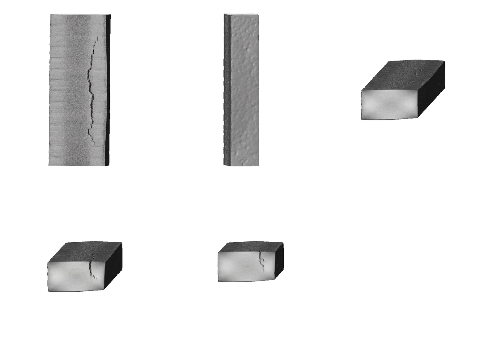
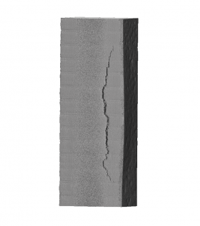
- Hover over a thumbnail and click on its overlaid description to see the animated GIF or a larger version of the image.
- MicroCT Scans for Industrial Materials
-
-
-
-
-
-
-
-